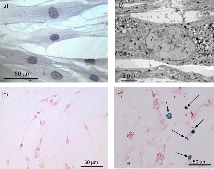
Fig 1. Prussian blue staining and TEM on FRDA fibroblasts.
a) Light microscopic image of healthy human fibroblasts; b) TEM image of human fibroblast; c) negative Prussian blue staining of human control fibroblasts; d) positive Prussian blue staining of fibroblasts from Friedreich’s ataxia (FRDA) patients. Iron-rich regions are clearly present in the FRDA fibroblast cells as bright blue regions (indicated with arrows), while fibroblast nuclei and cytoplasm have a red and pink color, respectively.

