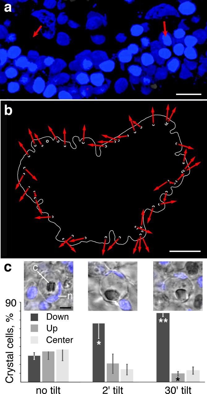
Fig 2. Orientation of crystal cells and position of crystal.
(a) Cup shaped nuclei of two crystal cells are apparent in an en face view of the edge of an animal stained with Hoechst nuclear dye. Orientations of the cups are indicated by red arrows. Oval-shaped nuclei of epithelial cells are tightly packed near the edge of the animal (below). Volume rendering of a series of confocal optical sections was created with Leica 3D imaging software. (b) Map of the positions and orientations of the cup-shaped nuclei of crystal cells. Outline traces the edge of the animal. Orientations of the openings of the cups are indicated by red arrows in mouths of the cups. Most cup openings are oriented towards the perimeter. (c) Effect of gravity on the positions of crystals inside crystal cells. Insets show the positions of crystals (c) inside their crystal cell in an animal on a horizontal surface (left), or tilted vertically for 2 min (center) or 30 min (right). Graphs compares the proportion of crystal cells with crystal shifted up, down or in the center under the three conditions. Error bars are SEM. *—p<0.05. **—p<0.005. Abbreviations: c–crystal; n–crystal cell nucleus. Scale bars: a– 5 μm; b– 100 μm; c– 2 μm (insets).

