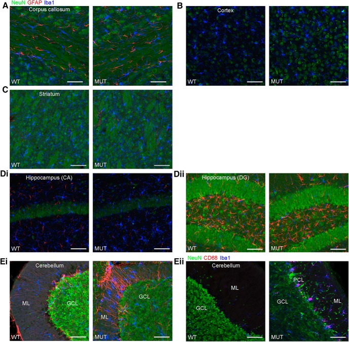
Figure 15.
Glial activation in the cerebellum of Nhe6-null mice at 2 mo of age. A–Eii, Representative images of 30-μm brain sections from 2-mo-old WT and MUT male littermate mice after triple immunohistochemical staining with antibodies against NeuN (green), a marker for neurons; GFAP (red), a marker for astrocytes (A–Ei), or CD68 (red), a marker for activated microglia (Eii); and Iba1 (blue), a marker for microglia. Coronal sections were used for CC (A), cortex (B), striatum (C), CA region of the hippocampus (Di), and DG region of the hippocampus (Dii), and sagittal sections were used for cerebellum (Eiand Eii). The amount of gliosis appears similar between MUT male mice and WT male littermates in CC, cortex, striatum, and hippocampus at 2 mo of age (A–Dii). However, gliosis in the cerebellum, especially in the ML, is apparent at this age in MUT male mice. This is reflected in the strong signals for GFAP and Iba1 (Ei) and CD68 and Iba1 (Eii), the latter of which is indicative of activated microglia. ML, molecular layer; PCL, Purkinje cell layer; GCL, granule cell layer. Scale bars, 50 μm.

