. 2017 Dec 21;81(1):80–87. doi: 10.4046/trd.2017.0108

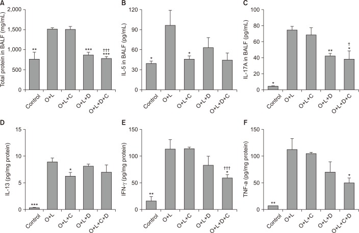
Figure 4. TH17 inflammation was attenuated by combination of macrolide and corticosteroid. Total protein, TH2 inflammation, TH17 inflammation, and proinflammatory cytokine in BALF analysis was increased after OVA+LPS challenge. (A) The number of total protein in BALF was decreased in DXM and CAM+DXM group. (B) IL-5 in BALF, marker of TH2 inflammation, was not significantly decreased in DXM or CAM+DXM group. (C) IL-17, marker of TH17 inflammation, was significantly decreased in DXM and CAM+DXM group. (D) IL-13, marker of TH2 inflammation, was evaluated in lung tissue. There was no significant decrease in DXM or CAM+DXM group. (E, F) Proinflammatory cytokine was decreased in combination of CAM+DXM and it was not decreased in monotherapy of CAM or DXM. O+L vs. all (*p<0.05, **p<0.01, ***p<0.001), O+L+C vs. O+L+D+C (p<0.05, †††p<0.001). BALF: bronchoalveolar lavage fluid; O: ovalbumin (OVA); L: lipopolysaccharide (LPS); C: clarithromycin (CAM); D: dexamethasone (DXM); IL: interleukin; IFN-γ: interferon γ; TNF-α: tumor necrosis factor α.

Figure 4