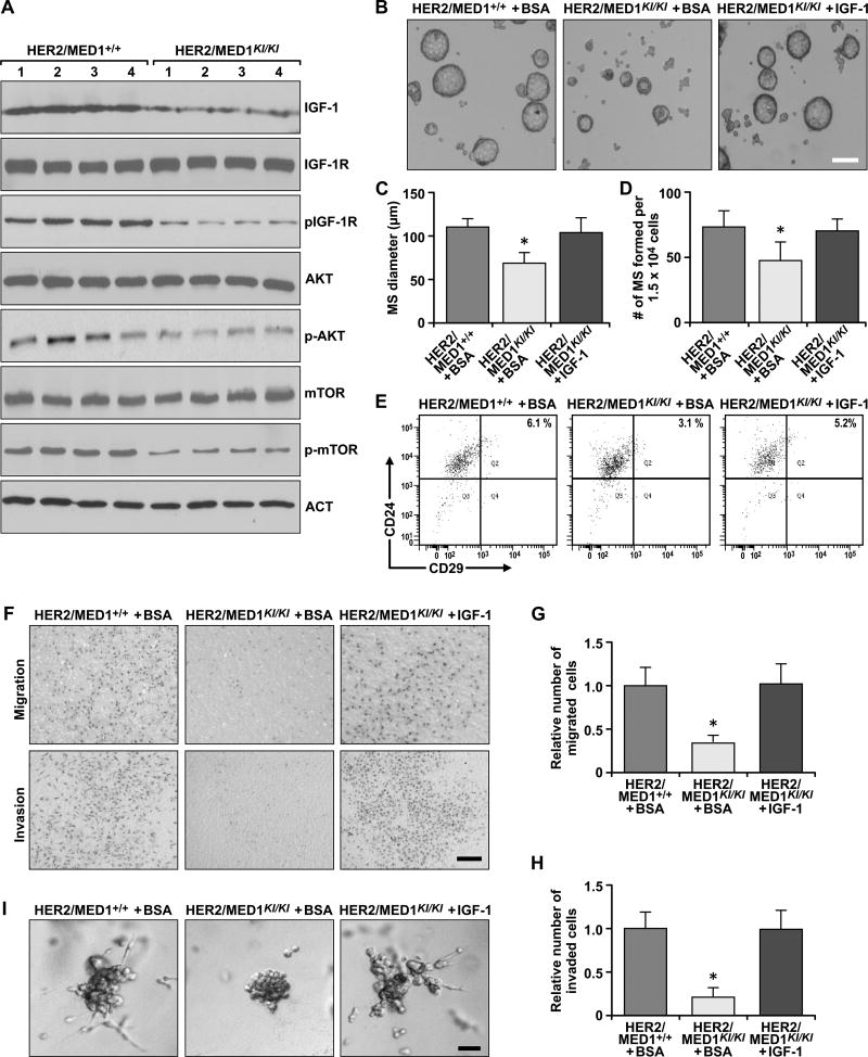
Fig 6. Exogenous IGF-1 rescues metastatic and mammosphere formation of MMTV-HER2/MED1KI/KI tumor cells in vitro.
(A) Immunoblotting analyses of the expression and status of IGF-1 and its downstream signaling pathway components in MMTV-HER2/MED1+/+ and MMTV-HER2/MED1KI/KI tumors. (B) Mammosphere formation assays using FACS sorted MMTV-HER2/MED1+/+ and MMTV-HER2/MED1KI/KI tumor cells in the presence of BSA or IGF-1. Bar = 100 µm. (C–D) Average diameters (C) and numbers (D) of mammospheres formed in (B). (E) Flow cytometry analyses of CD24+CD29hi CSCs in the mammospheres of (B). (F) Transwell assays using MMTV-HER2/MED1+/+ and MMTV-HER2/MED1KI/KI tumor cells in the presence of BSA or IGF-1. (G–H) Quantification of relative number of migrated (G) and invaded (H) cells in (F). (I) 3D culture of MMTV-HER2/MED1+/+ and MMTV-HER2/MED1KI/KI tumor cells in the presence of IGF-1 or BSA. Bar = 20 µm. The values are obtained in three independent experiments and shown as mean ± SD. *P < 0.05 or **P < 0.01.

