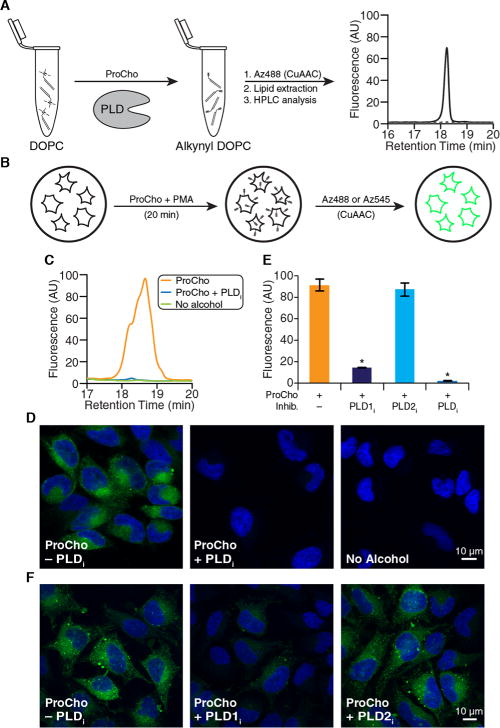
Figure 2.

ProCho can report on acutely stimulated PLD activity. (A) HPLC analysis of in vitro transphosphatidylation of dioleoylphosphatidylcholine (DOPC) with ProCho in the presence (solid line) or absence (dotted line) of A. hypogaea PLD, following CuAAC tagging with Az488. (B) Schematic for labeling cellular PLD activity. (C–F) HeLa cells were treated with PLD inhibitors [FIPI (750 nM), VU0359595 (250 nM), or VU0364739 (350 nM)] for 30 min, followed by ProCho (100 μM) and PMA (100 nM) for 20 min. (C and E) Lipid extracts were subjected to CuAAC with Az488 and HPLC analysis. (D and F) Cells were fixed, tagged with Az545 via CuAAC, and imaged by confocal microscopy: green for CuAAC-derived fluorescence and blue for DAPI. (E) Quantification (area under curve) of labeled extracts tagged with Az488 via CuAAC and analyzed by HPLC. n = 3; *p < 0.05. Error bars indicate the standard error of the mean. Abbreviations: PLDi, FIPI; PLD1i, VU0359595; PLD2i, VU0364739; PMA, phorbol 12-myristate 13-acetate.
