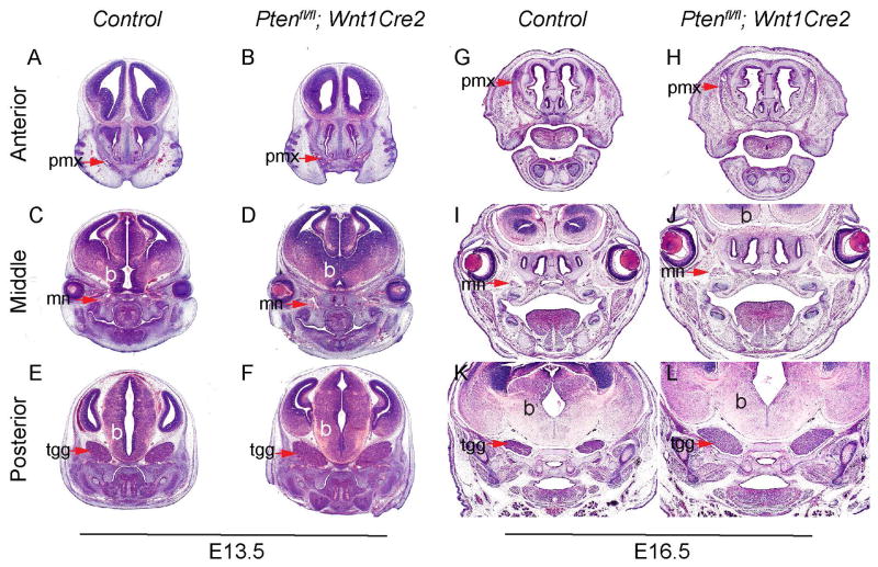
Fig 2. Ptenfl/fl; Wnt1Cre2 embryos exhibit overgrowth of craniofacial tissues.
H&E staining on frontal sections of littermate control and Ptenfl/fl; Wnt1Cre2 at E13.5 (A–F) and at E16.5 (G–L). Arrows point to pmx in A, B, G and H, to mn in C, D, I and J, and to tgg in E, F, K and L. b, brain; mn, maxillary nerve; pmx, premaxilla; tgg, trigeminal ganglion.

