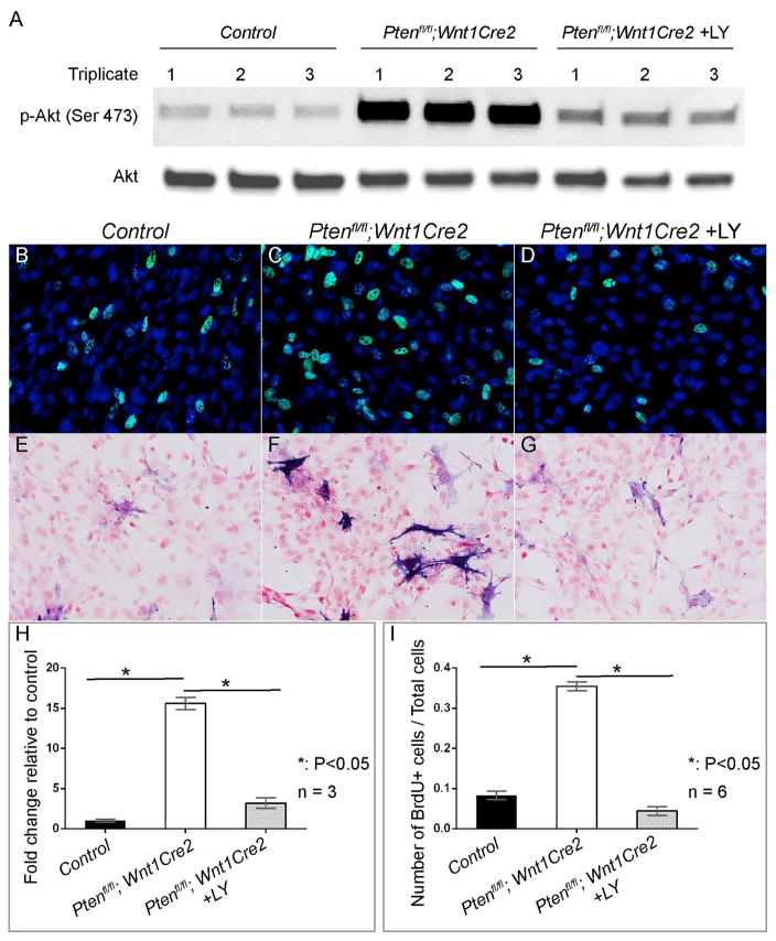
Fig 7. Inhibiting PI3K/AKT signaling rescues Pten mutant neural crest cells phenotype in vitro.
(A) Western blot of phospho-AKT and total AKT in primary pmx cells prepared from littermate control and Ptenfl/fl; Wnt1Cre2 embryos at E13.5. (B–D) BrdU labeling of primary pmx cells of littermate control (B), Ptenfl/fl; Wnt1Cre2 (C) and Ptenfl/fl; Wnt1Cre2 treated with 10uM LY for 30 minutes (D). BrdU+ cells are labeled with green fluorescence and total nuclei are stained with DAPI (blue). (E–G) AP staining of primary E13.5 pmx cells of littermate control (E), Ptenfl/fl; Wnt1Cre2 (F) and Ptenfl/fl; Wnt1Cre2 treated with 10uM LY for 8 hours (G). AP positive cells are blue and nuclei are counterstained with nuclear fast red. (H) Quantification and statistic analysis of western blot result of (A). (I) Quantification and statistic analysis of BrdU labeling results of (B–D).

