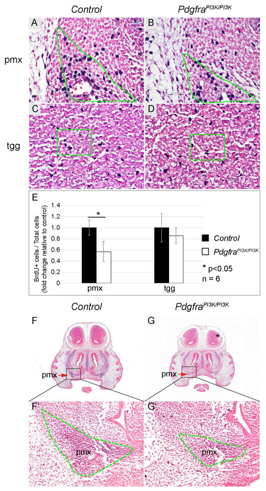
Fig 8. Attenuation of PI3K/AKT signaling affects NCCs proliferation and differentiation during craniofacial development.
(A–D) BrdU labeling on frontal sections of littermate control (A, C) and PdgfraPI3K/PI3K (B, D) embryos at E13.5, with a focus on the pmx (A, B) and tgg (C, D). Slides were counterstained with nuclear fast red to facilitate quantification. (E) Quantification and statistical analysis of percentile of BrdU+ cells in the control and PdgfraPI3K/PI3K tissues. The result is presented as fold change relative to the control. (F, G) AP-AB staining on frontal sections of littermate control (F) and PdgfraPI3K/PI3K (G) embryos at E13.5. (F′, G′) Magnification of defined area in F and G. pmx, premaxilla; tgg, trigeminal ganglion.

