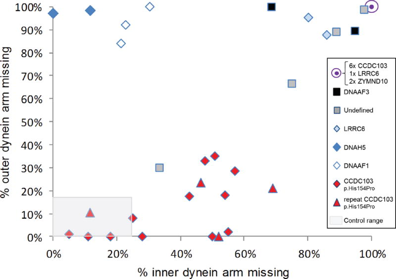
Figure 3. Quantitative transmission electron microscopy survey of inner and outer dynein arm loss in CCDC103 p.His154Pro patients versus a comparator group.

By surveying >100 cross sections in each patient sample we performed quantitative electron microscopy to determine the percentage of arm defects in cilia from individuals homozygous for the hypomorphic p.His154Pro CCDC103 mutation. Red diamonds and triangles indicate results from 16 CCDC103 p.His154Pro homozygote PCD patients, where triangles indicates the result of 4 repeat nasal brushings performed on patients marked by a diamond. The comparator group of 16 individuals with PCD, indicated by other symbols as shown, consists of 3 cases with DNAAF1 mutations (open diamonds), 2 DNAAF3 (black squares), 2 DNAH5 (dark blue diamonds), 3 LRRC6 (2 as light blue diamonds, 1 contained within the filled purple circle), 2 ZMYND10 (contained within the filled purple circle) and 4 cases in whom no mutations in known genes could be identified (grey squares). 6 CCDC103 p.His154Pro samples (27%) of p.His154Pro samples showed complete lack of both outer and inner dynein arms comparable to other gene mutations in the graph (purple circle). The grey shaded area represents normal range counts from >200 non PCD respiratory controls.30 Four CCDC103 p.His154Pro patients had counts within this normal range (one is a repeat sample (triangle) which showed similar data). Individuals with CCDC103 p.His154Pro mutation have a trend towards a distinctive pattern of partial loss of dynein arms that diverges from total dynein arm loss in the comparator group.
