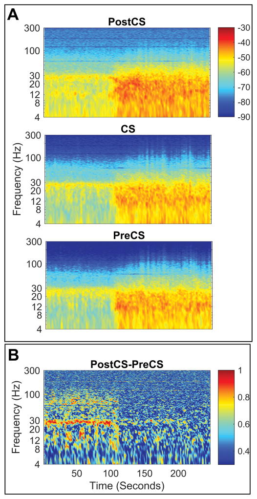
Figure 4.
High β power changes precedes changes in other bands in Parkinson disease group with proopfol anesthesia. (A-B) Time frequency representation of relative timing between changes in band power changes for different frequency bands for two representative subjects, highlighting similarity in relative pattern despite difference in timing of the changes. Time stamps are relative to the beginning of the recording (0 seconds) and Propofol was injected at T = 60 seconds.

