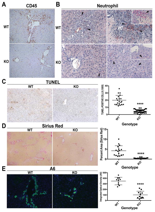
Figure 2. Decreased inflammation and fibrosis in KO following BDL.
(A) IHC shows fewer CD45+ inflammatory cells especially in the periportal region (100x). (B) Naphthol-ASD chloroacetate esterase staining shows fewer infiltrating neutrophils in KO after BDL (200x; inset 400x). (C) Reduced TUNEL-positive cells in KO after BDL (100x). (D) Greatly reduced fibrosis was seen in KO (50x). (E) IF for A6 shows notably less ADP in KO after BDL as compared to WT (100x). For C, 5 representative 100x images per animal (n=3 animals per group) were quantified; for D, 1–3 representative 50x images per animal (n=7 animals per group) were quanitified; for E, n=5 images per 2 representative animals per group were quantified. ****p<0.0001

