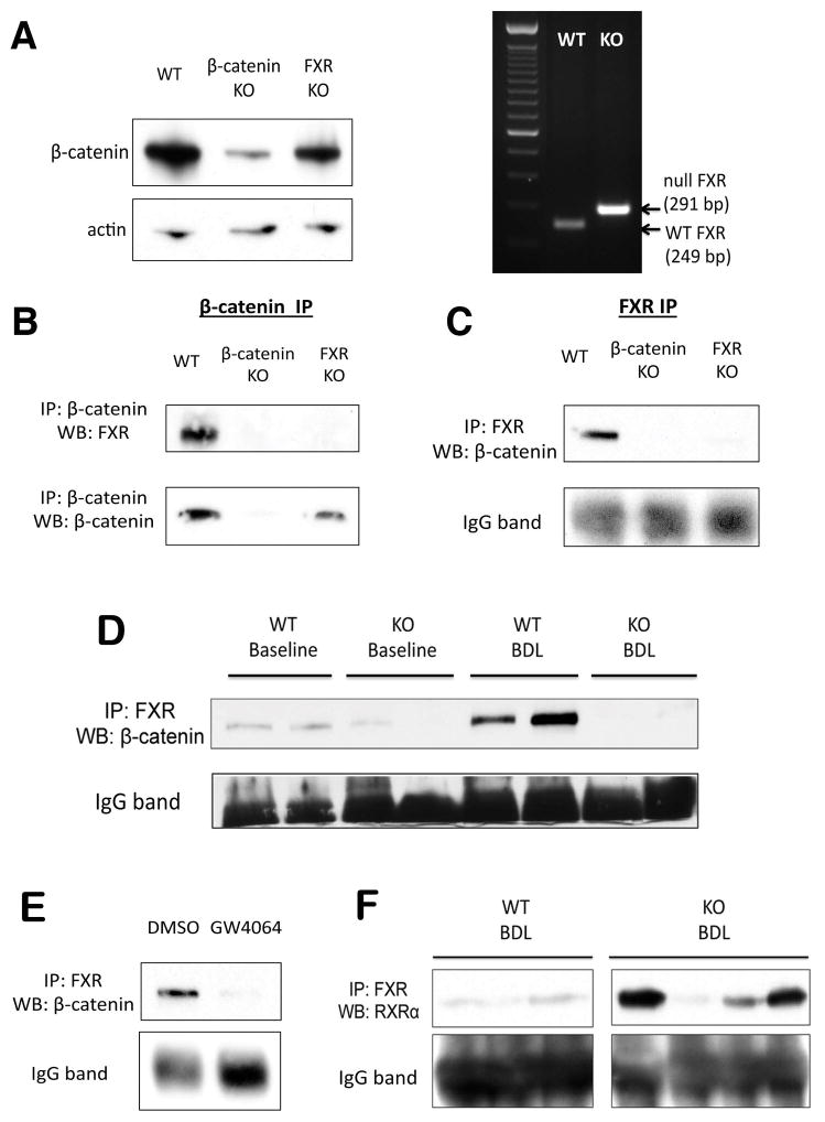
Figure 6. FXR and β-catenin form a complex in hepatocytes.
(A) WB for β-catenin confirms loss of β-catenin from β-catenin KO livers (right), and PCR of genomic DNA confirms deletion of FXR from FXR KO (left). (B) IP with β-catenin shows association with FXR only in WT livers but not in β-catenin KO or FXR KO. (C) Reverse IP with FXR antibody also demonstrates pull-down of β-catenin only in WT sample. (D) IP shows FXR/β-catenin association in WT livers is maintained after BDL. As expected, this association is decreased in KO. (E) Association between β-catenin and FXR decreases in the presence of FXR agonist GW4064 in Hep3B cells, as demonstrated by IP. (F) A notable increase in FXR/RXRα association is seen in most KO after BDL, as compared to WT after BDL.

