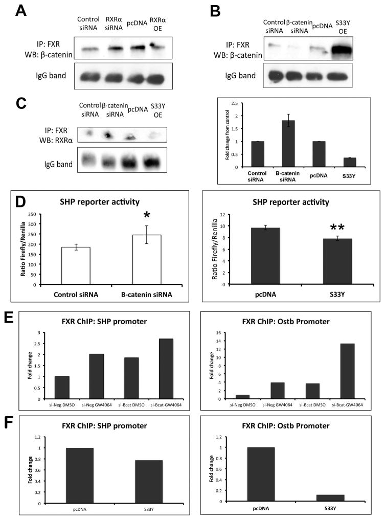
Figure 7. Manipulation of β-catenin and RXRα expression results in altered FXR/β-catenin association.
(A) IP shows that suppression of RXRα increases FXR/β-catenin, while overexpression of RXRα decreases FXR/β-catenin association. (B) Suppression of β-catenin decreases FXR/β-catenin association, while overexpression of β-catenin increases FXR/β-catenin association. (C) Left panel: Suppression of β-catenin increases FXR/RXRα association, while overexpression of β-catenin decreases FXR/RXRα association. Right panel: Densitometry confirms increased FXR/RXRα association in the absence of β-catenin and decreased FXR/RXRα when β-catenin is overexpressed (bars represent an average of n=2 independent experiments). (D) Left panel: Measurement of SHP reporter activity in the presence of β-catenin siRNA confirms increased activation of FXR in Hep3B cells. *p<0.05. Right panel: Expression of S33Y-mutated β-catenin in Hep3B cells resulted in notably lower activation of SHP reporter compared to control. **p<0.01. (E) ChIP assay demonstrates increased occupancy of FXR on the SHP and Ostβ promoters after GW4064 treatment; this increase also occurs following β-catenin suppression alone, and is further increased in the presence of GW4064. (F) Overexpression of β-catenin through transfection of S33Y plasmid led to a marked decrease in occupancy of the SHP and Ostβ promoters by FXR. ChIP data represents pooled samples from n≥3 plates per treatment group.

