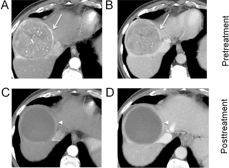
Fig. 3. Hepatocellular carcinoma (HCC) treated with drug-eluting bead transarterial chemoembolization (DEB-TACE).
Biopsy-proven HCC (arrows) in segment 8 showing arterial phase hyperenhancement (A), washout and capsule on portal venous phase (B). After DEB-TACE, no residual enhancement except for a thin rim of enhancement (arrowheads) is seen on arterial (C) and portal venous phases (D) (LR-TR Nonviable).

