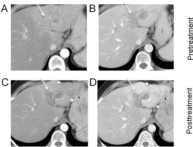
Fig. 7. Hepatocellular carcinoma (HCC) with tumor in vein (arrows), treated with transarterial radioembolization (TARE).
Biopsy-proven HCC in the left hepatic lobe with tumor in vein on arterial phase (A) and portal venous phase (B). One month after TARE, the tumor in vein demonstrates decreased enhancement, but hyperenhancement in the surrounding treated left lateral segment (arrowheads) is visible on both arterial phase (C) and portal venous phase (D). Vague areas of enhancement within the tumor in vein are equivocal for tumor viability (LR-TR Equivocal 4.4 cm).

