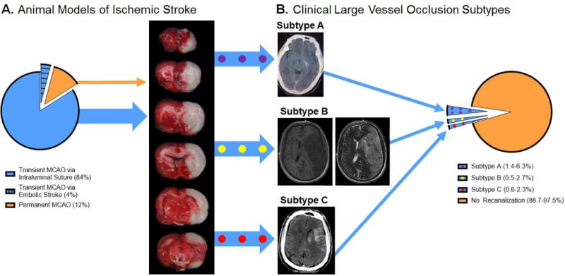
Fig 1. Disparity Between Cerebral Ischemia Animal Models and Clinical Stroke Subtypes.

A. Animal models of cerebral ischemia. Animal models of LVO include the transient MCAO and permanent MCAO models. Transient MCAO can be induced via an intraluminal suture or embolism. The intraluminal suture transient MCAO model (blue) accounted for 84% of the preclinical stroke studies in 2014 and 2015. Transient MCAO via embolic stroke (blue with black lines) accounted for 4% of the 2014 and 2015 experimental stroke studies. Permanent MCAO (orange) accounted for 12% of the 2014 and 2015 preclinical stroke studies. A representative image of a TTC-stained brain from a rat subjected to 2 hours of MCAO via intraluminal suture is shown. The red-stained tissue indicates healthy tissue. The white, unstained tissue indicates the infarction. This brain has an infarction volume equal to 26% of the whole brain (or 52% of the ipsilesional hemisphere). Transient MCAO can mimic clinical LVO patients of subtypes A (blue with purple dots), B (blue with yellow dots), and C (blue with red dots). B. Clinical distribution of LVO stroke patients. The number of patients with LVO is approximately 300,000 per year in the United States. Of these patients, 10,950–27,000 have vessel recanalization, which can be categorized using the large vessel recanalization subtypes A, B, and C. Of the LVO patients, 1.4–6.3% are subtype A (blue with purple circles), 0.5–2.7% are subtype B (blue with yellow circles), and 0.6–2.3% are subtype C (blue with red circles). The remaining large vessel stroke patients, 273,000–289,050 (88.7–97.5%), do not have vessel recanalization (orange). Representative images of each patient subtype are shown (subtype A: CT image, subtype B: T1 MRI (left) and T2 MRI (right), subtype C: CT).
