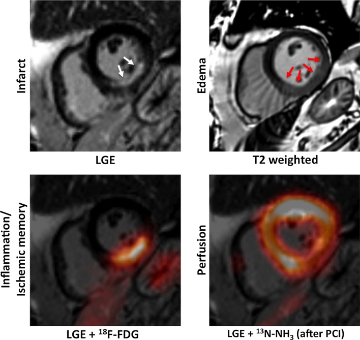
Fig. 8.
Multimodal characterization of the myocardial tissue after AMI using PET/MRI. Short-axis images of a patient who was imaged shortly after acute MI using simultaneous 18F-FDG and 13N-NH3 PET/MRI. Myocardial scarring can be imaged using LGE MRI (left column, top; white arrows pointing at subendocardial non-transmural infarction). The area of myocardial infarction is exceeded by the myocardial oedema imaged using T2-weighted sequences (right column, top; red arrows). Using fasting-heparin 18F-FDG-PET/MRI, the area of post-ischemic inflammation or ischemic memory can be assessed. After revascularization by percutaneous coronary intervention (PCI), only a slightly reduced perfusion of the inferior wall was observed in this patient [113]

