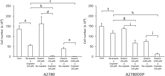
Figure 7.
Effect of a short time pre-exposure of erastin on proliferation of human ovary cell lines in the presence of cisplatin. A2780 (left panel) and A2780DDP (right panel) cells were seeded on 35 mm dishes (2 × 10⁵ cells/dish), cultured for 24 h, and exposed to 10 µM or 100 µM erastin for 5 min. After removing erastin from the medium, the cells were washed with 1 mL of fresh medium once and were allowed to grow in fresh medium in the presence or absence of 10 µM cisplatin. After culturing the cells for 48 h, the number of viable cells was counted by the trypan blue-exclusion method. Bars represent the mean of cell number ± S.D. (n = 10 for control of A2780 and A2780DDP; n = 4 for other conditions). P values were obtained by unpaired Student’s t test. Each small letters indicate statistical significance. P values are a = 2 × 10⁻³, b = 3 × 10⁻⁴, c = 4 × 10⁻⁴, d = 3 × 10⁻³, e = 2 × 10⁻³, f = 3 × 10⁻³, g = 1.5 × 10⁻³, h = 9 × 10⁻⁴, i = 2 × 10⁻³, j = 2 × 10⁻⁴.

