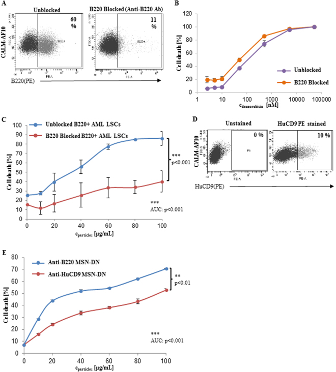
Figure 2.
(A) FACS dot plot demonstrating B220 positivity of the B220+ AML LSCs after blockage with the unlabeled anti-B220 antibody for 4 hours and subsequent staining with the anti- B220-PE conjugated antibody compared to an unblocked control (n = 3). Cell death of B220+ AML LSCs blocked by the anti-B220 antibody after incubation with different concentrations of (B) free daunorubicin (n = 3) or (C) with anti-B220 MSN-DN compared to unblocked cells (n = 3) (p = n.s. and p < 0.001, respectively). (D) Representative FACS dot plot showing expression of the CD9 antigen on B220+ AML LSCs (CALM-AF10 cells)(n = 3). (E) Percent death of B220+ AML LSCs induced by daunorubicin loaded MSNs at different particle concentrations after 24 hours incubation as indicated (n = 3)(p < 0.01).

