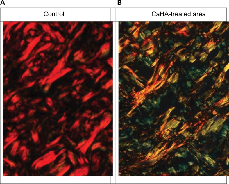
Figure 2.

Histological sections of the dermis stained with picrosirius red, observed under the microscope equipped with circularly polarized light.
Notes: (A) Control, collagen fibers appear mainly red. (B) Following CaHA injection, collagen fibers appear mainly green and yellow. Some fiber profiles appear orange.
