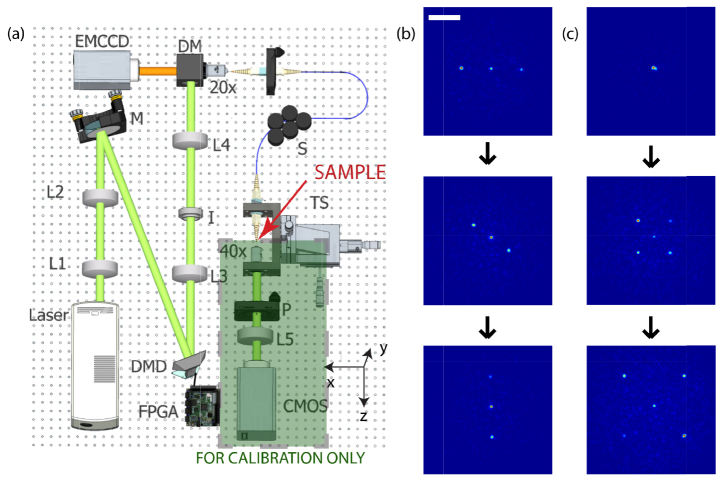
Fig. 2.
Fiber endoscope system and scanning capability. (a) Experimental apparatus to calibrate the MMF, measure the speckle statistics and perform fluorescence imaging. The laser beam illuminates L1,L2,L3,L4 and L5: lenses; I: Iris; S: Scrambler; TS: Translation stage; P: Polarizer. BS: Beam splitter. (b-c) Different dynamic patterns created at the distal tip consisting of three and five focus spots. In the left column three spots rotate clockwise, while in the right column five spots expand from the center of the fiber. A movie of the foci moving can be found in the supplementary material (Visualization 1 (216.6KB, MOV) and Visualization 2 (119.2KB, MOV) ). Scale bar is 25µm.

