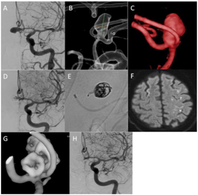
Figure 6.
A patient with an unruptured AComA aneurysm (a–c) was treated with 2 MEDs. There was good stasis of contrast in the aneurysm at the end of the procedure (d) however some persistent filling was seen. Upon removal of the microcatheter a single MED loop was displaced and could be seen in the parent artery (e). The patient was started on dual anti-platelet agents. He developed symptoms consistent with an ACA territory infarction and MRI demonstrated multiple small embolic infarctions within the ACA territory (f). He recovered completely and follow-up angiography showed complete occlusion of the aneurysm (g and h).
ACA: anterior cerebral artery; AComA: anterior communicating artery; MED: Medina Embolic Device; MRI: magnetic resonance imaging.

