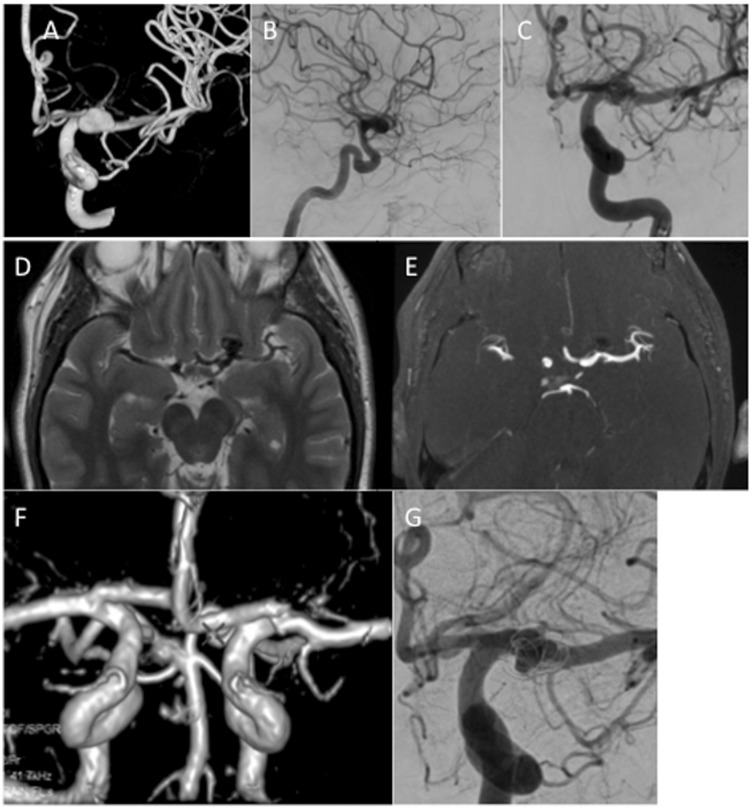
Figure 7.
A patient with an unruptured left ICA bifurcation aneurysm (a–c) was treated with a single MED. At initial follow up performed at 6 months axial T2-weighted MRI (d) showed a signal void in the aneurysm suggestive of continued filling however, on TOF MRA (e) there was no evidence flow which was confirmed on 3D reconstruction of the MRA data (f). On angiography, there was persistent filling of the aneurysm (mRRC IIIa). The discrepancy between the MRI and the angiographic data was thought to be due to a ‘Faraday cage’ like effect of the MED.
ICA: internal carotid artery; MED: Medina Embolic Device; MRA: Magnetic Resonance Angiography; MRI: magnetic resonance imaging; mRRC: modified Raymond–Roy occlusion classification; TOF: Time of Flight.

