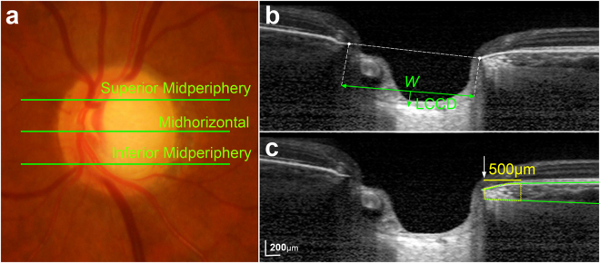
Figure 3.
Determination of the lamina cribrosa curvature index (LCCI) and juxtapapillary choroidal thickness (JPCT). (a) Sterescopic optic disc photograph image. (b,c) B-scan images obtained at superior midperiphery as shown in (a). (b) The LCCI was measured by dividing the lamina cribrosa (LC) curve depth (LCCD) within Bruch’s membrane (BM) opening by length of LC surface reference line (W), and then multiplying by 100. (c) Green solid lines indicate the upper and lower margins of the parapapillary choroid at the superior midperiphery, represented by BM and the choroidoscleral interface, respectively. The area of the juxtapapillary choroidal tissue within 500 μm from the border tissue of Elschnig was measured, and the mean was calculated by dividing the area by 500 μm.

