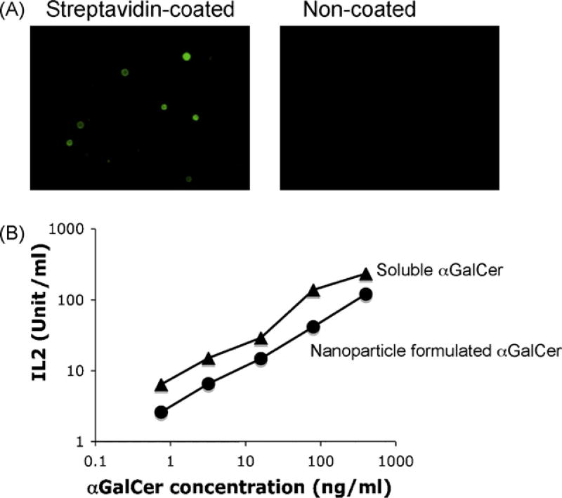
Fig. 1.
Presentation of nanoparticle formulated αGalCer by dendritic cells. (A) Strepatavidin coated nanoparticles were stained by biotin-FITC, and visualized by fluorescence microscope. Nanoparticles without streptavidin coating (non-coated) were stained as negative control. (B) Strepatvidin coated nanoparticles were conjugatedwith biotinylated αGalCer, and presented by mouse dendritic cells to stimulate NKT cells. Soluble form of biotinylated αGalCer were used as positive control. NKT cell activation was represented by their secretion of cytokine (IL2). Data were representative of three independent experiments: (▲) soluble biotinylated αGalCer and (●) nanoparticle conjugated αGalCer.

