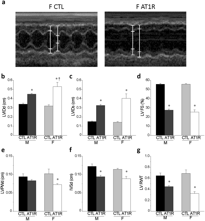
Figure 4.
Echocardiographic data reveal that female AT1R mice develop more severe left ventricular eccentric hypertrophy and dilation than AT1R males. (a) Typical examples of an M-mode echocardiography recordings in female CTL and AT1R mice. (b,c) The left ventricular internal diameter at end-diastole (LVIDd) and end-systole (LVIDs) were calculated from the echocardiography data obtained from CTL and AT1R mice of both sexes. (d) The LV fractional shortening representing the systolic function was calculated as follows: LV FS (%) = [(LVIDd − LVIDs)/LVIDd] × 100. (e,f) The echocardiographic data were also used to assess the LV posterior wall thickness (LVPWd) (e) and the interventricular septum (IVSd) (f) for each groups. (g) The relative wall thickness was determined as follows: LV RWT = (IVSd + LVPWd)/LVIDd. These data indicate that AT1R stimulation leads to more pronounced eccentric hypertrophy and dilation in females than males (*p < 0.005 vs. sex-matched CTL, †p = 0.05 vs. M AT1R) (M: CTL N = 7, AT1R N = 7; F: CTL N = 5, AT1R N = 8). Echocardiography values of male CTL and AT1R mice presented in this figure represent a subset of the data published in22.

