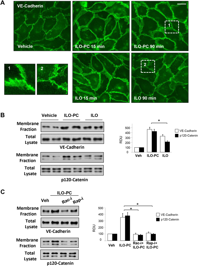
Figure 5.
Effects of ILO and ILO-PC on rapid and sustained enhancement of adherens junctions. (A) EC monolayers were incubated with 0.5 µM ILO or ILO-PC for 15 or 90 min. Increased areas of adherens junctions in response to agonist treatment were monitored by immunofluorescence staining of transmembrane adherens junction protein, VE-cadherin. Bar = 10 µm. Insets with higher magnification images show details of adherens junction structure. (B) Membrane accumulation of adherens junction proteins VE-cadherin and p120-catenin after 90-min stimulation with 0.5 µM ILO or ILO-PC detected by subcellular fractionation assay. (C) Membrane accumulation of VE-cadherin and p120-catenin caused by ILO or ILO-PC was analyzed in the presence or absence of Rac1 and Rap1 pharmacological inhibitors (30-min pre-treatment with 100 µM NSC23766 and 10 µM GGTI298, respectively). Probing of total lysates for VE-cadherin and p120-catenin was used as a normalization control. Bar graphs depict quantitative densitometry analysis of western blot data; n = 4; *p < 0.05. Data are expressed as mean ± SD. Biological replicates indicated by (n). Statistical significance by one-way analysis of variance (ANOVA) and Tukey’s post hoc multiple-comparison test.

