Figure 1.
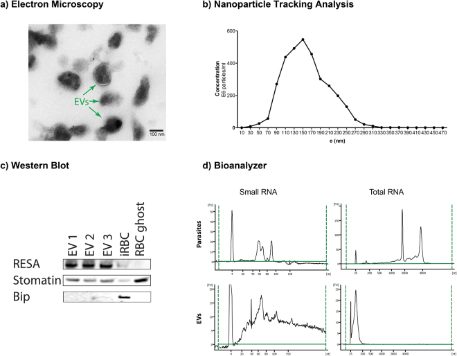
Extracellular vesicles isolated from P. falciparum carry RNA. (a) Transmission electron microscopy of EV preparation, purified EVs show individual EVs and a few clumps of varying sizes and intact lipid bilayers. The scale bar is 100 nm. (b) Representative particle size distribution from purified EVs. The concentration and size are determined by Nanoparticle Tracking Analysis. One representative experiment is shown. (c) Isolated EVs were confirmed by Western Blot for the enrichment of EV markers stomatin and RESA and negative for the endoplasmic reticulum Bip. (d) Total and Small bioanalyzer profiles of EV RNAs and total cell RNA show enrichment of small RNAs in EVs. One representative experiment is shown.

