Figure 3.
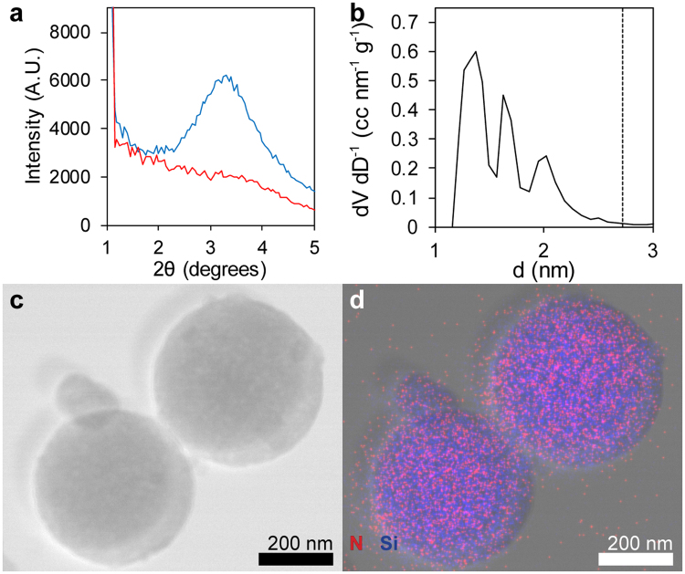
Physical characterization of OCT-MSNs demonstrates the relationship between templating drug molecule and MSN structure. The displayed XRD results (a) contain a low, broad diffraction peak typical of as synthesized OCT-templated MSNs (blue), as well as the lack of peak in particles synthesized below the OCT critical micelle concentration (red). DFT analysis (b) of N2 adsorption data shows 3 pore diameters (▬) approximately ½ the XRD d-spacing (▪▪▪). Scanning TEM (c) and overlaid EDX map (d) of OCT-MSNs confirms the presence of OCT with particles: Si signal emanates from the silica particles, while N (unique to the loaded OCT) signal is confined to the area of MSNs.

