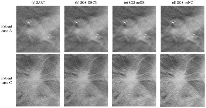
Fig. 9.
Comparison of four methods using human subject DBT images with invasive ductal carcinomas. The sizes of the image patches are 150 × 160 pixels (top row) and 300 × 360 pixels (bottom row). The SART method used 3 iterations. The parameters were β = 70, δ = 0.002/mm for the SQS-DBCN method, β = 40, δ = 0.002/mm for the SQS-noDB method, and β = 30, δ = 0.002/mm for the SQS-noNC method. All SQS methods were run for 10 iterations. All images are displayed with the same window width setting.

