Figure 1.
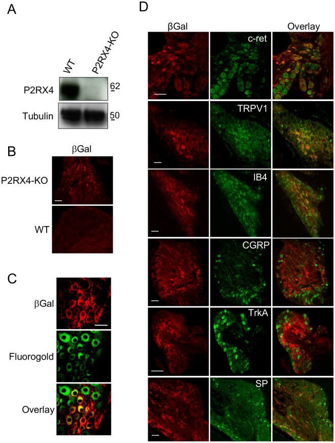
P2RX4 expression in Dorsal Root Ganglion neurons. (A) Representative cropped western blotting analysis of lumbar DRG extract indicates the presence of a 60 kDa band in P2RX4+/+ DRG that is absent in extracts from P2RX4−/− DRG. N = 3 independent experiments, n = 3 mice per genotype. (B) β-galactosidase staining was used as a surrogate marker for P2RX4 in DRG sections. Only P2RX4−/− neurons are immuno-positive for ßgal compared to sections from P2RX4+/+ DRG. Scale bar 100 µm. (C) DRG neurons that express ß-gal innervate paw skin. Fluoro-Gold B, a retrograde marker of neurons was injected in paw-skin and L5/L6 DRG were removed for staining a week later. βgal immunostaining reveals a population of neurons that are labeled for both ßgal and Fluoro-Gold B. Scale bar 50 µm. (D) In nociceptive neurons, ßgal is mainly expressed in c-ret-, TRPV1- and IB4-positive neurons, but scarcely in others populations (CGRP, TrkA or Substance P (SP) expressing cells). Representative images of DRG sections stained with βgal and makers of nociceptive neurons. Scale bars 50 µm.

