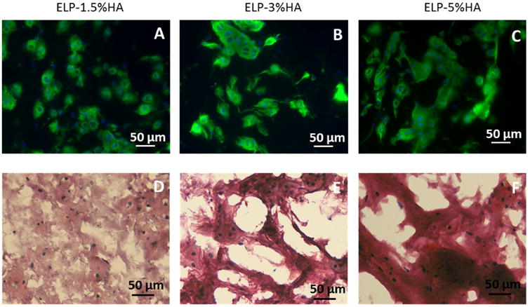
Figure 5.

The morphology of newly formed cartilage nodules (type II collagen and sGAG) by chondrocytes in ELP-HA hydrogels after 21 days of culture. (A-C) Immunostaining of type II collagen shows overall comparable amount of collagen II across the three groups, while the nodule size increases as HA increases; (D-F) Safranin O staining shows increasing HA concentration led to increased sulfated-glycosaminoglycan (sGAG) deposition. Scale bars: 50 μm.
