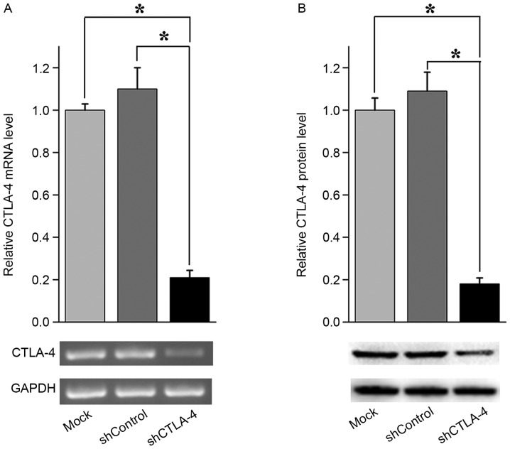
Figure 2.
CTLA-4 shRNA knocked down CTLA-4 expression in CIK cells. (A) Agarose gel images and qPCR analysis indicated that CTLA-4 mRNA expression was down-regulated in shCTLA-4 lentiviral particles transduced CIK cells compared with mock transduced CIK cells or shControl lentiviral particles transduced CIK cells. *P<0.01. CTLA-4 mRNA expression levels were normalized to GAPDH. CTLA-4 mRNA expression from shControl lentiviral particles transduced CIK cells and shCTLA-4 lentiviral particle-transduced CIK cells were normalized to 1 for mock transduced CIK cells. (B) Western blot analysis indicated that CTLA-4 protein expression was down-regulated in shCTLA-4 lentiviral particles transduced CIK cells compared with mock transduced CIK cells or shControl lentiviral particles transduced CIK cells. *P<0.01. CTLA-4 protein expression levels were normalized to GAPDH. CTLA-4 protein expression from shControl lentiviral particles transduced CIK cells and shCTLA-4 lentiviral particles transduced CIK cells were normalized to 1 for mock transduced CIK cells. The data presented are mean values from three independent experiments and plotted as the mean ± standard error of the mean. CTLA-4, cytotoxic T lymphocyte-associated antigen 4; CIK, cytokine-induced killer; PMBC, peripheral blood mononuclear cells; qPCR, quantitative polymerase chain reaction; sh, short hairpin.

