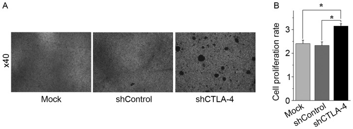
Figure 3.
Inhibition of CTLA-4 expression promoted CIK cell expansion in vitro. CIK cells on day 10 were transduced with shCTLA-4 lentiviral particles, shControl lentiviral particles, or mock transduced. A total of 96 h after cell transduction, CIK cells were harvested and counted to evaluate cell expansion efficiency. (A) CIK cells mock transduced (left panel), transduced with shControl lentiviral particles (middle panel), or transduced with shCTLA-4 lentiviral particles (right panel). Magnification, ×40. (B) Compared with mock transduced CIK cells or shControl lentiviral particles transduced CIK cells, shCTLA-4 lentiviral particles transduced CIK cells exhibited a higher expansion rate. *P<0.01. Each experiment was performed three times. The data are plotted as the mean ± standard error of the mean. CTLA-4, cytotoxic T lymphocyte-associated antigen 4; CIK, cytokine-induced killer; PMBC, peripheral blood mononuclear cells; qPCR, quantitative polymerase chain reaction; sh, short hairpin.

