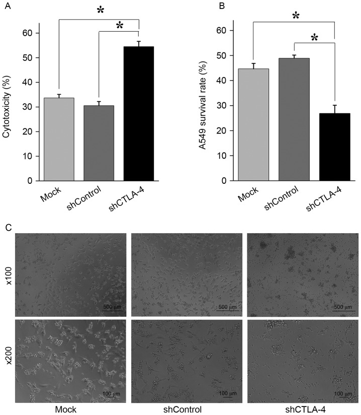
Figure 4.
The inhibition of CTLA-4 expression increased cytotoxicity of CIK cells against A549 cells. (A) Determined by CytoTox 96 Non-Radioactive Cytotoxicity Assay, compared with CIK cells mock transduced or transduced with shControl lentiviral particles, CIK cells transduced with shCTLA-4 lentiviral particles exhibited significantly higher cytotoxicity against A549 cells. *P<0.001. (B) A549 cells were co-cultured with CIK cells for 24 h before cell counting. Compared with the number of viable A549 cells co-cultured with CIK cells mock transduced or transduced with shControl lentiviral particles, the number of viable A549 cells co-cultured with CIK cells transduced with shCTLA-4 lentiviral particles were significantly lower. *P<0.01. (C) Representative figures of A549 cells co-cultured with CIK cells mock transduced (left panel), transduced with shControl lentiviral particles (middle panel), and transduced with shCTLA-4 lentiviral particles (right panel) for 24 h. Each experiment was performed three times. The data were plotted as the mean ± standard error of the mean. CTLA-4, cytotoxic T lymphocyte-associated antigen 4; CIK, cytokine-induced killer; PMBC, peripheral blood mononuclear cells; qPCR, quantitative polymerase chain reaction; sh, short hairpin.

