
Figure 2. DL-PDMP inhibits the proliferation of primary aneuploidy cells.
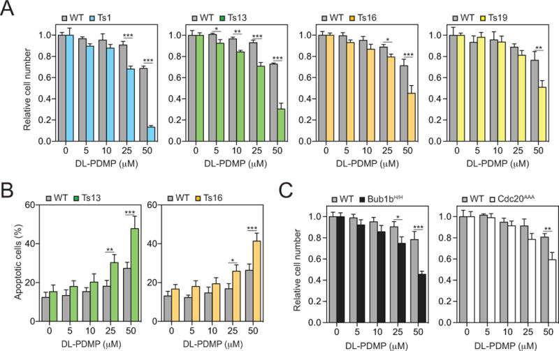
(A) Euploid wild-type (grey bars) and trisomic MEFs (colored bars) were treated with the indicated concentrations of DL-PDMP. Cell number was determined after 72 hours. In each experiment, aneuploid MEFs were compared to euploid littermate control MEFs. To specifically assess the effects of DL-PDMP on cell proliferation we normalized cell number of drug treated cells to that of the same cells treated with vehicle only. This was necessary because aneuploid cells proliferate poorly.
(B) Percentage of annexin V-FITC positive, PI negative cells was determined 24 hours after DL-PDMP treatment.
(C) Euploid wild-type (grey bars), BUB1bH/H cells (black bars) and CDC20AAA/AAA cells (open bars) were treated with the indicated concentrations of DL-PDMP. Cell number was determined after 72 hours. In each experiment, aneuploid MEFs were compared to euploid littermate control MEFs. To specifically assess the effects of DL-PDMP on cell proliferation we normalized cell number of drug treated cells to that of the same cells treated with vehicle only. This was necessary because aneuploid cells proliferate poorly.
The data are shown as the mean ± standard deviation. *P<0.05, **P<0.01, ***P<0.001, t test.