
Figure 4. Up-regulation of BCL6 by glucose deprivation works with secondary re-stimulation and is specific for BCL6.
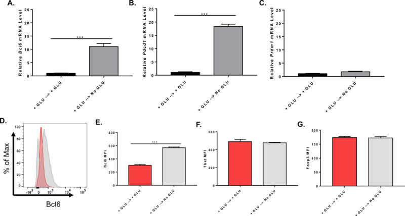
Naïve CD4+ T cells isolated from C57BL/6 mice were cultured with complete media for 48 h on anti-CD3 and anti-CD28 coated plate, cells were collected and washed once with PBS and cultured under complete (GLU+) or glucose (GLU-) deprivation medium for another 48 h on anti-CD3 and anti-CD28 coated plate. Cells were harvested for total RNA preparation or flow cytometric staining. (A) Relative mRNA expression was determined by QPCR. Bcl6 gene expression from isolated naïve CD4+ T cells cultured under complete or glucose deprivation medium during re-stimulation (n=3, mean ± SEM). (B) Pdcd1 gene expression from isolated naïve CD4+ T cells cultured under complete or glucose deprivation medium during re-stimulation (n=3, mean ± SEM). (C) Prdm1 gene expression from isolated naïve CD4+ T cells cultured under complete or glucose deprivation medium during re-stimulation (n=3, mean ± SEM). (D) and (E) Bcl6 MFI of naïve CD4+ T cells cultured under complete or glucose deprivation medium during re-stimulation (n=3, mean ± SEM). (F) Tbet MFI of naïve CD4+ T cells cultured under complete or glucose deprivation medium during re-stimulation (n=3, mean ± SEM). (G) Foxp3 MFI of naïve CD4+ T cells cultured under complete or glucose deprivation medium during re-stimulation (n=3, mean ± SEM). *p < 0.05, ***p < 0.001 (t test). Data are representative of two independent experiments with similar results.