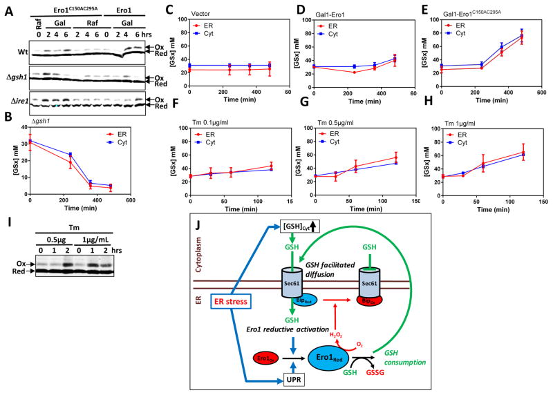
Fig. 6. Reciprocal interplay between the ER import of GSH and Ero1 activation.
(A) Kar2 oxidation in Wt, Δgsh1, and Δire1 expressing Ero1C150AC295A or Ero1 from a GAL1 promoter, as indicated, grown in raffinose or galactose medium for the indicated time and processed as in Figure 5B, C. (B–H) Corrected cytosolic (Cyt) (TotCell x 4.5) and ER (ER) [GSx] in Δgsh1 incubated during the indicated time in medium lacking GSH (B); in HGT1 cells carrying an empty vector (C), Gal1-ERO1 (D), or Gal1-ERO1C150AC295A (E), switched from raffinose to galactose medium at time 0 (C–E); in Wt cells incubated with 0.1 (F), 0.5 (G) or 1 μg/mL (H) tunicamycin (Tm) for the indicated time (F–H). (I) Wt cells incubated with 0.5 or 1 μg/mL tunicamycin (Tm) for the indicated time were processed for Kar2 oxidation as in Figure 5B, C. The 1- and 2-h 0.5μg/ml Tm samples contain ½ and ¼ the lysates amount relative to untreated (0 h), respectively, and the 1- and 2-h 1 μg/mL samples ¼ and 1/6 relative to 0 h. Values in B–H are the mean of three independent biological replicas ± standard deviation (s.d.). (J) Proposed model. ER stress stimulates both [GSH]Cyt increase and UPR-dependent Ero1 de novo synthesis. [GSH]Cyt increase stimulates ER facilitated diffusion of GSH, which promotes Ero1 reductive activation, and further stimulates GSH import by GSH oxidation to GSSG. UPR-dependent Ero1 level increase is critical for allowing active Ero1-dependent H2O2 production to reach the threshold levels required to oxidize Kar2, which inhibits GSH import, in turn allowing Ero1 activity to cease.

