Figure 2.

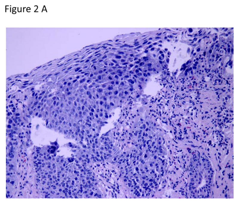
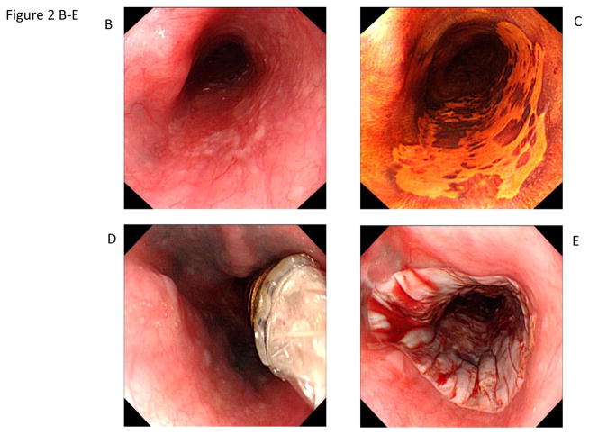
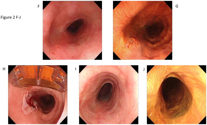
Circumferential and focal radiofrequency ablation of a 3-cm long flat-type early squamous cell neoplasia with high-grade intraepithelial neoplasia, treated with Lugol’s-10J/cm2-10J/cm2-no cleaning between ablation passes (circumferential technique Group D). The patient achieved a complete response (absence of moderate-grade intraepithelial neoplasia, high-grade intraepithelial neoplasia, and esophageal squamous cell carcinoma in the treatment area) at the 12-month primary outcome.
A. Photomicrograph of a pre-treatment esophageal biopsy specimen demonstrating HGIN (hematoxylin and eosin [H&E]; original magnification × 200).
B. Pretreatment white-light endoscopy image showing a reddish colored area from 4 o’clock to 7 o’clock.
C. Corresponding image with Lugol’s chromoendoscopy demonstrating a flat-type unstained lesion; biopsy samples showed HGIN.
D. Circumferential ablation catheter placed in the esophagus before the first ablation pass.
E. Appearance of the mucosa after the first circumferential ablation pass.
F. 3-month visit. White-light endoscopy image showing the treatment area.
G. Corresponding image with Lugol’s chromoendoscopy demonstrating an unstained lesion at 8 o’clock.
H. Appearance of the mucosa immediately after focal ablation of the unstained lesion; the ablation catheter can be seen at the top of the endoscopic image.
I–J.12-month primary endpoint visit. White-light endoscopy and Lugol’s high-resolution chromoendoscopy images demonstrate no evidence of residual squamous neoplasia. Biopsies confirmed complete response.


