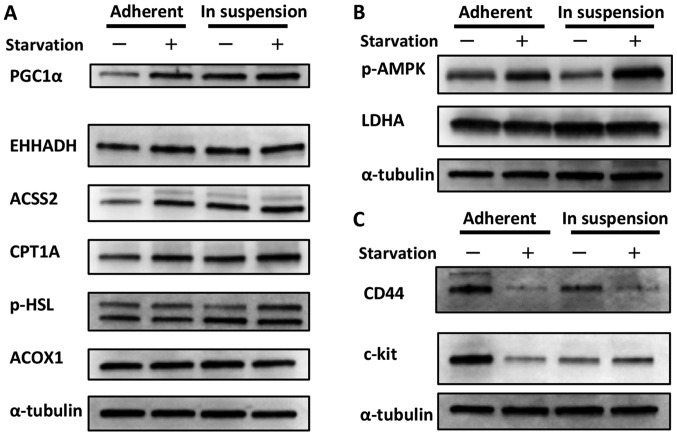
Figure 1.
Change in the metabolic status and CSC marker expression of cells in adherent plates vs. suspension. (A) Change in lipid metabolism. Cells cultured in suspension showed increased expression of PGC1α, which is thought to be a sensor for lipid metabolism, compared with cells cultured in adherent plates. Additionally, the expression of some types of enzymes related to fatty acid metabolism were also increased, although the level of increase depended on the enzyme. The expressions of these enzymes were increased in cells cultured in adherent plates when they were exposed to starvation; however, the cells cultured in suspension showed less sensitivity to starvation. This could be interpreted to indicate that cells in suspension already utilize fatty acid metabolism and maintain a relative advantage against starvation. (B) Change in glycolysis. The phosphorylation of AMPK, a marker for glycolysis, was decreased in cells cultured in suspension compared with cells cultured in adherent plates. The expression of LDHA, the indicator for glycolysis, was nearly unchanged between the two groups. Although the phosphorylation of AMPK was increased in cells cultured in suspension and exposed to starvation, it is possible that these cells were preparing for upcoming nourishment. (C) Change in CSC marker expression. Cells cultured in suspension showed decreased expression of CD44 and c-kit compared with cells cultured in adherent plates. Interestingly, the response during starvation differed between the two culture groups. While the expression of CD44 was decreased during starvation in cells cultured in both suspension and adherent plates, the expression of c-kit increased in cells cultured in suspension but decreased in cells cultured in adherent plates.

