Figure 1.
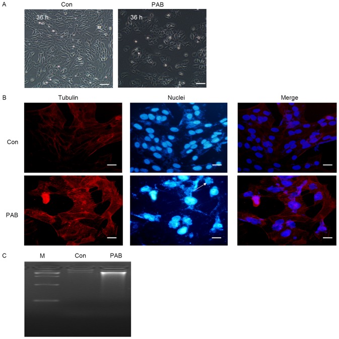
Apoptosis was not induced in MRC5 cells following treatment with PAB. (A) Morphological changes were observed in PAB-treated and control cells using phase contrast microscopy. Scale bar, 30 µm. (B) Cells were stained with phalloidin-tetramethylrhodamine B isothiocyanate and Hoechst 33258, and imaged with fluorescence microscopy, to determine morphological changes in the PAB-treated and control cells at 36 h. Arrows indicate multinuclear cells. Scale bar, 60 µm. (C) No fragmentation of chromosomal DNA was observed in PAB-treated cells at 36 h. All images are representative of three independent experiments. PAB, pseudolaric acid B; Con, control; M, marker.

