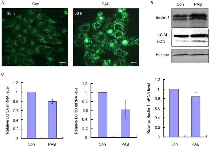
Figure 2.
PAB induced autophagy in MRC5 cells. (A) Treatment with PAB increased the number of monodansylcadaverine-stained dots detected in fluorescence microscopy. Scale bar, 90 µm. (B) Expression of LC 3 and Beclin-1. A western blot was performed at 36 h after PAB treatment. Histone H3 is included as a loading control. (C) At 36 h after PAB treatment, intracellular LC 3A, LC 3B and Beclin-1 mRNA levels were detected. The results, as mean ± standard deviation, were standardized to the expression of GAPDH mRNA and normalized to the control group. The results are representative of three independent experiments. PAB, pseudolaric acid B; LC 3, light chain 3; Con, control.

