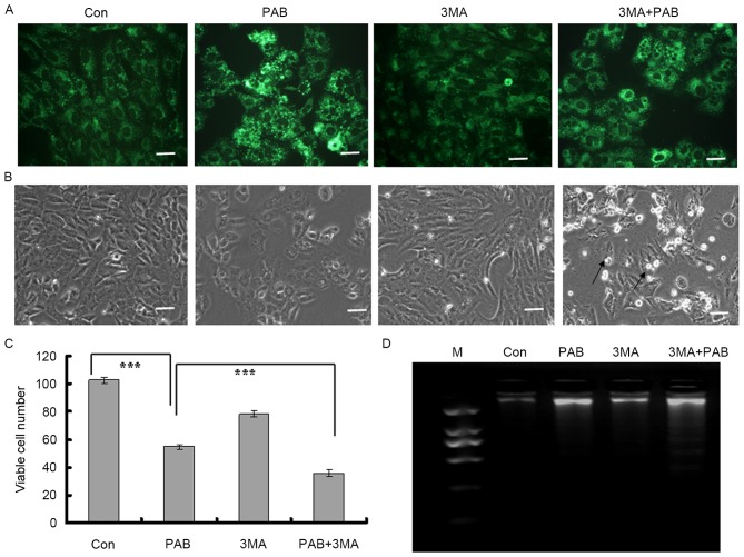
Figure 3.
Inhibiting autophagy promoted apoptosis in MRC5 cells subsequent to PAB treatment. (A) At 36 h after PAB and/or 3MA treatment, the number of monodansylcadaverine-stained dots was observed with fluorescence microscopy. Scale bar, 45 µm. (B) At 36 h after PAB and/or 3MA treatment, the morphological changes of MRC5 cells were observed with phase contrast microscopy. Arrows indicate the cells undergoing cell death. Scale bar, 30 µm. (C) At 36 h after PAB and/or 3MA treatment the cell number was counted with trypan blue staining and imaged via light microscopy. The data are the means ± standard deviation of 3 independent experiments. ***P<0.001. (D) Induction of chromosomal DNA fragmentation in cells treated with PAB and 3MA. PAB, pseudolaric acid B; 3MA, 3-methyladenine; Con, control; M, marker.

