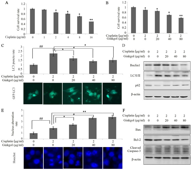
Figure 2.
Effects of ginkgol C17:1 on cisplatin-induced autophagy and apoptosis in HepG2 cells. (A) Following treatment with cisplatin (0, 1, 2, 4, 8 and 16 µg/ml) for 24 h, HepG2 cell viability was determined via an MTT assay. (B) Cell viability was detected by MTT assay following treatment with cisplatin (2 µg/ml) and ginkgol C17:1 (0, 20, 40 and 80 µg/ml) for 24 h. Following co-treatment with cisplatin and ginkgol C17:1, LC3 autophagosomes were detected by an (C) immunofluorescence assay (magnification, ×200) and the expression of Beclin-1, LC3I/II and p62 were analyzed by (D) a western blot assay. Under the same conditions, the morphology of HepG2 nuclei was observed by (E) immunofluorescence microscopy (magnification, ×200) staining with Hoechst 33342 and the expression of cleaved caspase-3, Bax and Bcl-2 were analyzed by (F) western blot analysis. Data are presented as the mean ± standard deviation from three independent experiments. *P<0.05 and **P<0.01 vs. control group, and ##P<0.01. LC3, light chain 3; Bcl-2, B-cell lymphoma 2; Bax, Bcl-2-associated X protein; GFP, green fluorescent protein.

