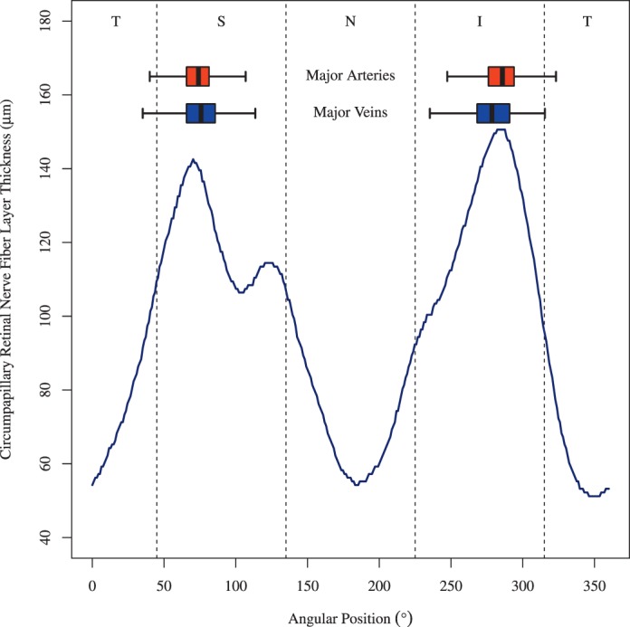
Figure 2.

BV position distribution over all eyes on the Cirrus standard diagnostic circle with 1.73 mm radius. As a spatial reference, the Cirrus cpRNFLT norm was added (blue line). Here, the norm for the age of 52 years is shown. The absolute cpRNFLT values decrease with age, but the peak of Cirrus norms remains at the same location according to the Cirrus age model. Boxplots: distributions of BV locations (box: first to third quartile; central mark: median; yellow point: mean; whiskers: fifth to 95th percentile). Dark red box: artery locations, dark blue box: vein locations. T, temporal; S, superior; N, nasal; I, inferior with respect to the ONH.
