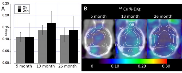
Figure 3. Whole brain regional 64Cu uptake at different ages assessed using longitudinal PET imaging.

(A) Whole brain 64Cu uptake (mean ± SD %ID/g) at 5, 13 and 26 months of age acquired at 2h and 24h post oral administration of the 64CuCl2 PET tracer. (B) Transaxial images showing regional 64Cu radioactivity (mean ± SD %ID/g) acquired 24h PO at 5, 13 and 26 months of age. Regions were shown for cerebellum (CB), Thalamus (T), olfactory bulb (OB), posterior cortex (PC), and frontal cortex (FC).
