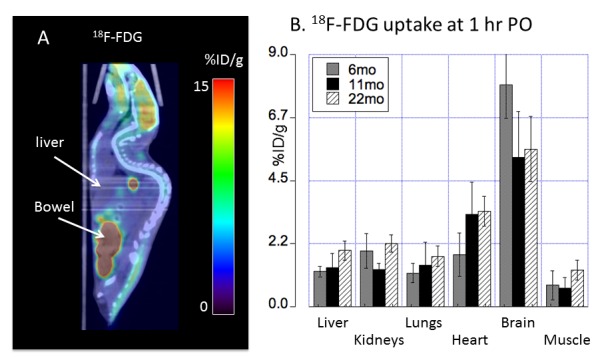
Figure 4. Biodistribution of 18F-FDG radioactivity (mean ± SD %ID/g) in C57BL/6 mice orally administered with 18F-FDG by PET/CT imaging.

(A) At 1h PO, prominent 18F-FDG radioactivity in the brain and gastrointestinal tracts was visualized on PET/CT images. Residual 18F-FDG was noted in oral cavity. (B) PET quantitative analysis demonstrated 18F-FDG tissue distribution across different ages (6, 11 and 22 months of age) with 18F-FDG uptake in the brain of middle and old age lower than 18F-FDG uptake in the brain of young adult C57BL/6 mice. Error bars represent SD; %ID/g: percentage of injected dose per gram tissue; PO: post oral administration of the tracer.
