Figure 3.

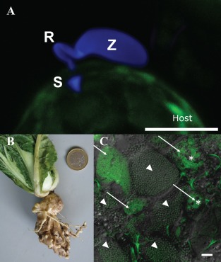
Phytomyxid infection and clubroot. (A) Phytomyxean parasites infect their host via a specialized extrusosome, called a ‘Rohr (R) and Stachel (S)’. The image shows a zoospore (Z) of the phagomyxid Maullinia ectocarpii infecting a female gametophyte of Macrocystis pyrifera (host). The M. ectocarpii spore was stained with calcofluor white and the host is visible via autofluorescence. Bar, 5 µm. (B) Clubroot symptoms on Chinese cabbage. (C) Laser scanning micrograph of Plasmodiophora brassicae resting spores (arrowheads) and plasmodia (arrows) in clubroot tissue. Plasmodia of different ages can be distinguished by the presence of typical vacuoles (asterisks), which disappear when the plasmodia start to differentiate into resting spores. Overlay of a light microscopic image and the signal of a Plasmodiophora‐specific fluorescence in situ hybridization (FISH) probe (green: excitation, 488 nm; emission, 510–550 nm). Bar, 20 µm.
