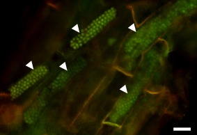
Figure 5.

Resting spores of Polymyxa graminis in Poa sp. Resting spores are arranged in typical, long and cylindrical cytosori (arrowheads). The sample was stained with acridine orange, showing the nuclei of the fully developed resting spores. Epifluorescence micrograph obtained using blue excitation with long‐pass emission (Nikon B‐2A filter) allowing for the detection of DNA. Bar, 20 µm.
